Knee Muscle Anatomy Mri : Posteromedial Corner Injury Of The Knee Radsource : Scroll through the structures to understand the anatomy.. The muscles of the knee include the quadriceps, hamstrings, and the muscles of the calf. 1 november 2002 mri anatomy of the knee and shoulder james y. Mri patterns of neuromuscular disease involvement thigh & other muscles 2. Rubin da, kettering jm, towers jd, britton ca: Tendons attach the muscles to each other.
Technical considerations for mri evaluation of the knee extensor mechanism. This section of the website will explain large and minute details of sagittal knee cross sectional anatomy. Click on the links to show each structure. Support the body in an upright position without the need for muscles to work. Song, uc san francisco msiv gillian lieberman md.
Stanford Msk Mri Atlas C 2020 from xrayhead.com Each anatomical structure was labeled interactively. Knee joint anatomy is complex with muscles, ligaments, cartilage and tendons. Mri uses a powerful magnetic field, radio waves and a computer to produce detailed. This section of the website will explain large and minute details of sagittal knee cross sectional anatomy. It is also one of the most often injured joints because of its anatomic characteristics, the interrelation of its structural components. 4, infrapatellar fat pad of hoffa. Anatomy of peritoneum and mesentery. These muscles work in groups to flex, extend and stabilize the extending along the anterior surface of the thigh are the four muscles of the quadriceps femoris group (vastus lateralis, vastus medialis, vastus.
The knee joint is one of the largest and most complex joints in the body.
Please email baodo at stanford.edu. Mri for evaluating knee pain in older patients: Knee joint anatomy is complex with muscles, ligaments, cartilage and tendons. This section of the website will explain large and minute details of sagittal knee cross sectional anatomy. Mr arthrogram knee loose osteochondral lesion. Want to learn more about it? Find out how the different structures fit together in our knee diagram the knee joint is the largest and one of the most complex joints in the human body. 4, infrapatellar fat pad of hoffa. Mr imaging of knees having isolated and combined ligament injuries. Click now to learn more about the bones, muscles, and soft tissues of these regions at leg and knee anatomy: Stanford msk mri atlas has served over 1,000,000 pages to users in over 100 countries. Scroll through the structures to understand the anatomy. This mri knee cross sectional anatomy tool is absolutely free to use.
Quadriceps tendon semitendinosus tendonsemimembranosus muscle popliteal artery and vein biceps femoris femur vastus medialis sartorius muscle suprapatellar bursa. 4, infrapatellar fat pad of hoffa. The journal of musculoskeletal medicine. Click on the links to show each structure. Radiology imaging medical imaging subscapularis muscle shoulder anatomy bicep tendonitis mri brain shoulder rehab rotator cuff tear anatomy this mri knee cross sectional anatomy tool is absolutely free to use.
Figure 12 From Normal Mr Imaging Anatomy Of The Knee Semantic Scholar from d3i71xaburhd42.cloudfront.net Injuries of the patellofemoral joint. An understanding of normal anatomy and biomechanics of the knee extensor mechanism is necessary to comprehend the imaging of extensor mechanism injuries. Free access interactive and dynamic anatomical atlas. Learn anatomy using a full pacs! 4, infrapatellar fat pad of hoffa. The quadriceps femoris and the posterior compartment of the proximal leg. The knee joint is one of the largest and most complex joints in the body. Helps to lower and raise the body.
Support the body in an upright position without the need for muscles to work.
Mr imaging of knees having isolated and combined ligament injuries. The knee is designed to fulfill a number of functions: Involved early gray = muscle: This mri knee cross sectional anatomy tool is absolutely free to use. Rubin da, kettering jm, towers jd, britton ca: This section of the website will explain large and minute details of sagittal knee. 12 photos of the knee muscle anatomy mri. The knee joint is one of the largest and most complex joints in the body. Musculoskeletal radiology south texas radiology group. Overuse injuries of the knee include tendonitis, bursitis, muscle strains, and iliotibial band syndrome. Each anatomical structure was labeled interactively. Click now to learn more about the bones, muscles, and soft tissues of these regions at leg and knee anatomy: Magnetic resonance imaging (mri) interpretation of the knee is often a daunting challenge to the student or physician in training.
Find out how the different structures fit together in our knee diagram the knee joint is the largest and one of the most complex joints in the human body. Tendons attach the muscles to each other. This mri knee cross sectional anatomy tool is absolutely free to use. Helps to lower and raise the body. Mr imaging of knees having isolated and combined ligament injuries.
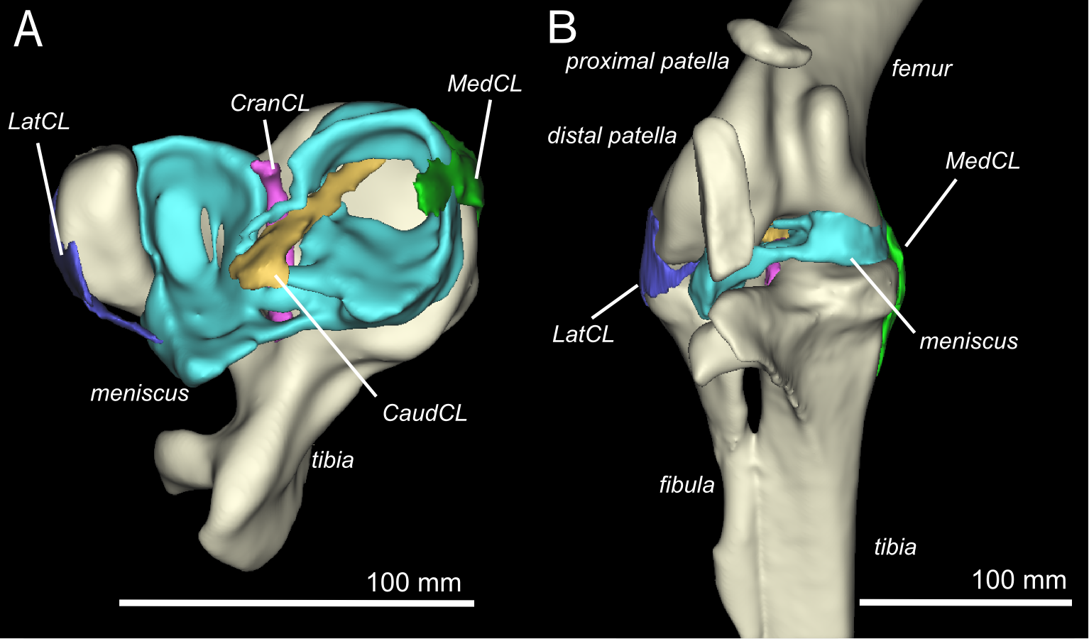
Three Dimensional Anatomy Of The Ostrich Struthio Camelus Knee Joint Peerj from dfzljdn9uc3pi.cloudfront.net Click now to learn more about the bones, muscles, and soft tissues of these regions at leg and knee anatomy: There are various muscles that control movement, ligaments that. They are attached to the femur (thighbone), tibia (shinbone), and fibula (calf bone) by fibrous tissues called ligaments. Musculoskeletal radiology south texas radiology group. Find out how the different structures fit together in our knee diagram the knee joint is the largest and one of the most complex joints in the human body. The muscles that affect the knee's movement run along the thigh and calf. Scroll through the structures to understand the anatomy. Rubin da, kettering jm, towers jd, britton ca:
Functional anatomy of the shoulder complex malcolm peat the shoulder complex, together with other joint and muscle mechanisms of the upper limb.
Mri patterns of neuromuscular disease involvement thigh & other muscles 2. Rubin da, kettering jm, towers jd, britton ca: Mri uses a powerful magnetic field, radio waves and a computer to produce detailed. Click on the links to show each structure. Each anatomical structure was labeled interactively. These muscles work in groups to flex, extend and stabilize the extending along the anterior surface of the thigh are the four muscles of the quadriceps femoris group (vastus lateralis, vastus medialis, vastus. Knee anatomy francesc malagelada jordi vega pau golanó the knee is the largest joint in the human body and one of the most complex from a functional point of view. Free access interactive and dynamic anatomical atlas. The muscles of the knee include the quadriceps, hamstrings, and the muscles of the calf. Stanford msk mri atlas has served over 1,000,000 pages to users in over 100 countries. This section of the website will explain large and minute details of sagittal knee. Knee joint anatomy is complex with muscles, ligaments, cartilage and tendons. Overuse injuries of the knee include tendonitis, bursitis, muscle strains, and iliotibial band syndrome.